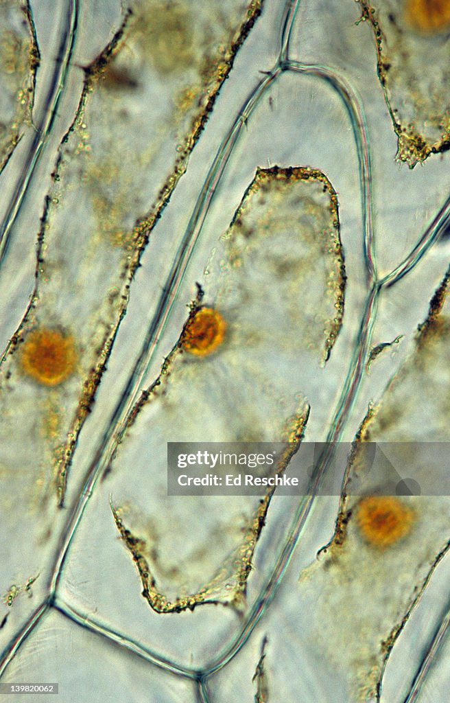
Plasmolysis. Onion Cells, Epidermis. 100x at 35mm. Onion cells were exposed to a hypertonic solution, & the protoplast lost water, shrunk & separated from cell wall. Shows: cell wall, nucleus, cytoplasm & cell membrane. Iodine stain. - stock photo

Get this image in a variety of framing options at Photos.com.
PURCHASE A LICENSE
All Royalty-Free licenses include global use rights, comprehensive protection, simple pricing with volume discounts available
€300.00
EUR
Getty ImagesPlasmolysis Onion Cells Epidermis 100x At 35mm Onion Cells Were Exposed To A Hypertonic Solution The Protoplast Lost Water Shrunk Separated From Cell Wall Shows Cell Wall Nucleus Cytoplasm Cell Membrane Iodine Stain High-Res Stock Photo Download premium, authentic Plasmolysis. Onion Cells, Epidermis. 100x at 35mm. Onion cells were exposed to a hypertonic solution, & the protoplast lost water, shrunk & separated from cell wall. Shows: cell wall, nucleus, cytoplasm & cell membrane. Iodine stain. stock photos from 51łÔąĎÍř Explore similar high-resolution stock photos in our expansive visual catalogue.Product #:139820062
 Download premium, authentic Plasmolysis. Onion Cells, Epidermis. 100x at 35mm. Onion cells were exposed to a hypertonic solution, & the protoplast lost water, shrunk & separated from cell wall. Shows: cell wall, nucleus, cytoplasm & cell membrane. Iodine stain. stock photos from 51łÔąĎÍř Explore similar high-resolution stock photos in our expansive visual catalogue.Product #:139820062
€300€40
Getty Images
In stockDETAILS
Credit:
51łÔąĎÍř #:
139820062
License type:
Collection:
Stone
Max file size:
1866 x 2911 px (6.22 x 9.70 in) - 300 dpi - 4 MB
Upload date:
Release info:
No release required
Categories:
- Biological Cell,
- Onion,
- Iodine,
- Backgrounds,
- Botany,
- Brightly Lit,
- Cell Membrane,
- Close-up,
- Color Image,
- Cytoplasm,
- Distorted,
- Full Frame,
- Hypertonic Solution,
- Immunofluorescent Photomicrograph,
- Iodine Stain,
- Leaf Epidermis,
- Living Organism,
- Magnification,
- Muscular Contraction,
- Natural Condition,
- No People,
- Nucleus,
- Pattern,
- Photography,
- Protoplasm,
- Research,
- Science,
- Scientific Micrograph,
- Scrutiny,
- Vertical,