Peripheral nerve, TEM - stock photo
Peripheral nerve. Coloured transmission electron micrograph (TEM) of a section through a small peripheral nerve. Myelin( blue) is an insulating fatty layer that surrounds the myelinated nerve fibres (pink), increasing the speed at which nerve impulses travel. It is formed when Schwann cells (yellow) wrap around the fibre, depositing layers of myelin between each coil. Unmyelinated nerve fibres are also present. An outer sheath or perineurium surrounds the connective tissue endoneurium that in turn surrounds the nerve fibres and Schwann cells. Magnification: x1500, when printed 10 centimetres wide.

Get this image in a variety of framing options at Photos.com.
PURCHASE A LICENSE
All Royalty-Free licenses include global use rights, comprehensive protection, simple pricing with volume discounts available
€300.00
EUR
Getty ImagesPeripheral Nerve Tem High-Res Stock Photo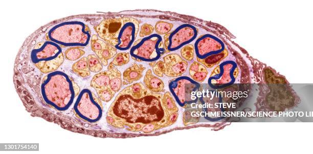 Download premium, authentic Peripheral nerve, TEM stock photos from 51łÔąĎÍř Explore similar high-resolution stock photos in our expansive visual catalogue.Product #:1301754140
Download premium, authentic Peripheral nerve, TEM stock photos from 51łÔąĎÍř Explore similar high-resolution stock photos in our expansive visual catalogue.Product #:1301754140€300€40
Getty Images
In stockDETAILS
51łÔąĎÍř #:
1301754140
License type:
Collection:
Science Photo Library
Max file size:
6096 x 3003 px (20.32 x 10.01 in) - 300 dpi - 4 MB
Upload date:
Location:
United Kingdom
Release info:
No release required
Categories: