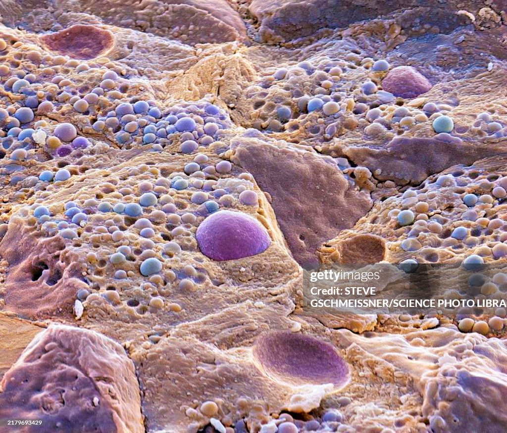
Pancreas tissue, SEM - stock photo
Pancreas tissue. Coloured scanning electron micrograph (SEM) of fractured exocrine pancreas tissue, showing numerous acinar cells, containing secretory zymogen granules. The freeze fracture has revealed larger cell nuclei (purple) and the depressions they sit in. Digestive enzymes are secreted in the zymogen granules, and passed to the small intestine through the pancreatic ducts. Magnification: x1600 when printed 10 centimetres wide

Get this image in a variety of framing options at Photos.com.
PURCHASE A LICENSE
All Royalty-Free licenses include global use rights, comprehensive protection, simple pricing with volume discounts available
€300.00
EUR
Getty ImagesPancreas Tissue Sem High-Res Stock Photo Download premium, authentic Pancreas tissue, SEM stock photos from 51łÔąĎÍř Explore similar high-resolution stock photos in our expansive visual catalogue.Product #:2179693429
 Download premium, authentic Pancreas tissue, SEM stock photos from 51łÔąĎÍř Explore similar high-resolution stock photos in our expansive visual catalogue.Product #:2179693429
€300€40
Getty Images
In stockDETAILS
51łÔąĎÍř #:
2179693429
License type:
Collection:
Science Photo Library
Max file size:
4572 x 3911 px (15.24 x 13.04 in) - 300 dpi - 10 MB
Upload date:
Location:
United Kingdom
Release info:
No release required
Categories: