TMICROSCOPIC ANATOMY (HISTOLOGY) of TRANSITIONAL EPITHELIUM (STRETCHED STATE), URINARY BLADDER, 250X - stock photo
MICROSCOPIC ANATOMY (HISTOLOGY) of TRANSITIONAL EPITHELIUM (Stretched state), URINARY BLADDER, 250X. Transitional Epithelium (found lining the bladder and ureter) consists of several layers of cells that can change their shape when the organ is stretched. The cells here assume a flattened shape when the bladder is stretched (when filling with urine) as opposed to a plumper shape when not stretched. This image also shows the muscular layer of the bladder--dark blue showing nuclei of smooth muscle cells. Below the muscular layer (called detrusor muscle), is connective tissue with a blood vessel filled with red blood cells (reddish appearance).

Get this image in a variety of framing options at Photos.com.
PURCHASE A LICENSE
All Royalty-Free licenses include global use rights, comprehensive protection, simple pricing with volume discounts available
€300.00
EUR
Getty ImagesTmicroscopic Anatomy Urinary Bladder 250x High-Res Stock Photo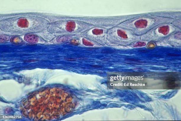 Download premium, authentic TMICROSCOPIC ANATOMY (HISTOLOGY) of TRANSITIONAL EPITHELIUM (STRETCHED STATE), URINARY BLADDER, 250X stock photos from 51łÔąĎÍř Explore similar high-resolution stock photos in our expansive visual catalogue.Product #:1369962756
Download premium, authentic TMICROSCOPIC ANATOMY (HISTOLOGY) of TRANSITIONAL EPITHELIUM (STRETCHED STATE), URINARY BLADDER, 250X stock photos from 51łÔąĎÍř Explore similar high-resolution stock photos in our expansive visual catalogue.Product #:1369962756€300€40
Getty Images
In stockDETAILS
Credit:
51łÔąĎÍř #:
1369962756
License type:
Collection:
Photodisc
Max file size:
5212 x 3495 px (17.37 x 11.65 in) - 300 dpi - 21 MB
Upload date:
Location:
United States
Release info:
No release required
Categories: