Neurone, SEM - stock photo
Neurone. Coloured scanning electron micrograph (SEM) of a PC12 neurone in culture.The PC12 cell line, developed from a pheochromocytoma tumor of the rat adrenal medulla, has become a premiere model for the study of neuronal differentiation. When treated in culture with nerve growth factor, PC12 cells stop dividing, elaborate processes, become electrically excitable, and will make synapses with appropriate muscle cells in culture. Magnification: x4000 when printed 10 centimetres wide

Get this image in a variety of framing options at Photos.com.
PURCHASE A LICENSE
All Royalty-Free licenses include global use rights, comprehensive protection, simple pricing with volume discounts available
€300.00
EUR
Getty ImagesNeurone Sem High-Res Stock Photo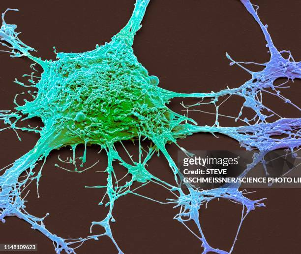 Download premium, authentic Neurone, SEM stock photos from 51łÔąĎÍř Explore similar high-resolution stock photos in our expansive visual catalogue.Product #:1148109623
Download premium, authentic Neurone, SEM stock photos from 51łÔąĎÍř Explore similar high-resolution stock photos in our expansive visual catalogue.Product #:1148109623€300€40
Getty Images
In stockDETAILS
51łÔąĎÍř #:
1148109623
License type:
Collection:
Science Photo Library
Max file size:
4572 x 3851 px (15.24 x 12.84 in) - 300 dpi - 5 MB
Upload date:
Location:
United Kingdom
Release info:
No release required
Categories:
- Brain Tumour,
- Human Nervous System,
- Biological Cell,
- Pheochromocytoma,
- Nerve Cell,
- SEM,
- Adrenal Medulla,
- Anatomy,
- Biology,
- Color Image,
- Color Manipulation,
- Colors,
- Connection,
- Dendrite,
- Educational Subject,
- Healthy Lifestyle,
- Horizontal,
- Human Body Part,
- Human Brain,
- Human Internal Organ,
- Organization,
- Photography,
- Physiology,
- Scanning Electron Microscope,
- Synapse,
- UK,