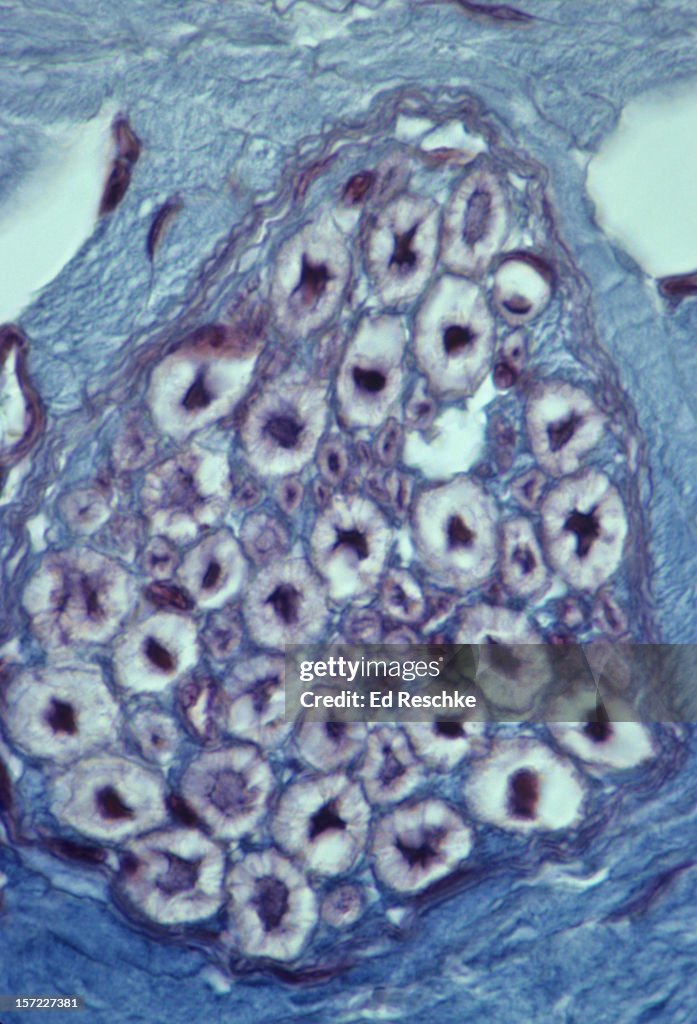
Myelinated Nerve Fibers--nerve in cross section - stock photo
Myelinated Nerve Fibers--nerve in cross section, magnification 250x. This image shows: nerve fibers (dark), myelin (white), endoneurium (blue, connective tissue), perineurium, and a single fasicle (a bundle of nerve fibers) within the nerve. masson stain

Get this image in a variety of framing options at Photos.com.
PURCHASE A LICENSE
All Royalty-Free licenses include global use rights, comprehensive protection, simple pricing with volume discounts available
€300.00
EUR
Getty ImagesMyelinated Nerve Fibersnerve In Cross Section High-Res Stock Photo Download premium, authentic Myelinated Nerve Fibers--nerve in cross section stock photos from 51łÔąĎÍř Explore similar high-resolution stock photos in our expansive visual catalogue.Product #:157227381
 Download premium, authentic Myelinated Nerve Fibers--nerve in cross section stock photos from 51łÔąĎÍř Explore similar high-resolution stock photos in our expansive visual catalogue.Product #:157227381
€300€40
Getty Images
In stockDETAILS
Credit:
51łÔąĎÍř #:
157227381
License type:
Collection:
Stone
Max file size:
3518 x 5166 px (11.73 x 17.22 in) - 300 dpi - 17 MB
Upload date:
Location:
United States
Release info:
No release required
Categories: