Immunofluorescent photomicrograph,Microscopic photograph of a professionally prepared slide of liver tissue. The liver is divided histologically into lobules. Organs samples, Histological examination, histopathology on the microscope - stock photo
Education anatomy and phistopathology of Tongue under the microscopic in laboratory.

Get this image in a variety of framing options at Photos.com.
PURCHASE A LICENSE
All Royalty-Free licenses include global use rights, comprehensive protection, simple pricing with volume discounts available
€300.00
EUR
Getty ImagesImmunofluorescent Photomicrograph Microscopic Photograph Of A Professionally Prepared Slide Of Liver Tissue The Liver Is Divided Histologically Into Lobules Organs Samples Histological Examination Histopathology On The Microscope High-Res Stock Photo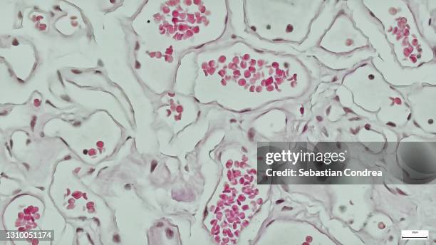 Download premium, authentic Immunofluorescent photomicrograph,Microscopic photograph of a professionally prepared slide of liver tissue. The liver is divided histologically into lobules. Organs samples, Histological examination, histopathology on the microscope stock photos from 51łÔąĎÍř Explore similar high-resolution stock photos in our expansive visual catalogue.Product #:1310051174
Download premium, authentic Immunofluorescent photomicrograph,Microscopic photograph of a professionally prepared slide of liver tissue. The liver is divided histologically into lobules. Organs samples, Histological examination, histopathology on the microscope stock photos from 51łÔąĎÍř Explore similar high-resolution stock photos in our expansive visual catalogue.Product #:1310051174€300€40
Getty Images
In stockDETAILS
Credit:
51łÔąĎÍř #:
1310051174
License type:
Collection:
Moment
Max file size:
3840 x 2160 px (12.80 x 7.20 in) - 300 dpi - 3 MB
Upload date:
Location:
United States
Release info:
No release required
Categories:
- White Blood Cell,
- Blood,
- Microscope,
- Blood Cell,
- Liver - Organ,
- Backgrounds,
- Hepatocyte,
- Tissue - Anatomy,
- Color Image,
- Magnification,
- Trombone,
- Abstract,
- Anatomy,
- Biological Cell,
- Biology,
- Biopsy,
- Cardiovascular System,
- Condition,
- Desktop PC,
- Education,
- Healthcare And Medicine,
- High-scale Magnification,
- Histology,
- Horizontal,
- Human Body Part,
- Human Cell,
- Human Digestive System,
- Human Internal Organ,
- Human Liver,
- Human Tissue,
- Human Vein,
- Illness,
- Immunofluorescent Photomicrograph,
- In Bloom,
- Light Micrograph,
- Medical Exam,
- Medicine,
- Microbiology,
- Microscope Slide,
- Natural Pattern,
- No People,
- Order,
- Pattern,
- Photography,
- Physiology,
- Research,
- Reticular Fiber,
- Science,
- Scientific Micrograph,
- Technology,
- USA,