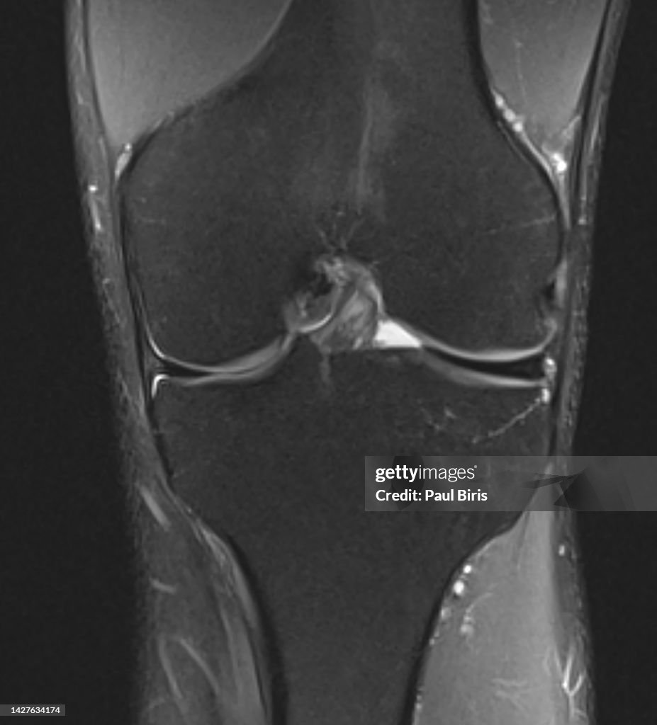
Discoid lateral meniscus seen on coronal PD FatSat MRI image - stock photo
Discoid menisci are anatomical variants that have a body that is too wide, usually affecting the lateral meniscus. They are incidentally found in 3-5% of knee MRI examinations

Get this image in a variety of framing options at Photos.com.
PURCHASE A LICENSE
All Royalty-Free licenses include global use rights, comprehensive protection, simple pricing with volume discounts available
€300.00
EUR
Getty ImagesDiscoid Lateral Meniscus Seen On Coronal Pd Fatsat Mri Image High-Res Stock Photo Download premium, authentic Discoid lateral meniscus seen on coronal PD FatSat MRI image stock photos from 51łÔąĎÍř Explore similar high-resolution stock photos in our expansive visual catalogue.Product #:1427634174
 Download premium, authentic Discoid lateral meniscus seen on coronal PD FatSat MRI image stock photos from 51łÔąĎÍř Explore similar high-resolution stock photos in our expansive visual catalogue.Product #:1427634174
€300€40
Getty Images
In stockDETAILS
Credit:
51łÔąĎÍř #:
1427634174
License type:
Collection:
Moment
Max file size:
6729 x 7429 px (22.43 x 24.76 in) - 300 dpi - 4 MB
Upload date:
Location:
Romania
Release info:
No release required
Categories:
- Analyzing,
- Anatomy,
- Black And White,
- Bone,
- Broken,
- Care,
- Commuter,
- Data,
- Diagnostic Medical Tool,
- Doctor,
- Examining,
- Expertise,
- Healthcare And Medicine,
- Hospital,
- Human Body Part,
- Human Bone,
- Human Joint,
- Human Knee,
- Human Tissue,
- Illness,
- Joint - Body Part,
- Knee,
- Laboratory,
- Leg,
- Limb - Body Part,
- MRI Scan,
- Magnet,
- Medical Clinic,
- Medical Exam,
- Medical Research,
- Medical Scan,
- Medical Scanner,
- Medical Test,
- Meniscus,
- Modern,
- No People,
- Orthopedic Surgeon,
- Orthopedist,
- Patient,
- Photography,
- Physical Injury,
- Radiologist,
- Romania,
- Science,
- Scientific Imaging Technique,
- Sport,
- Surgery,
- Technology,
- Tomography,
- Torn,
- Vertical,