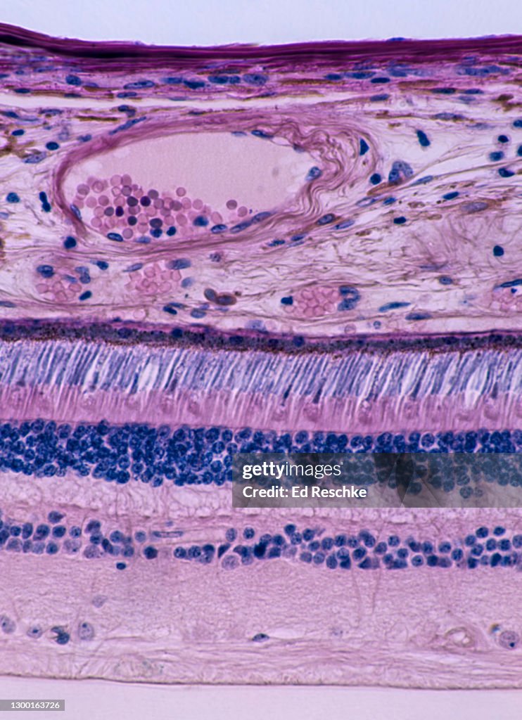
RETINA, ANATOMY--LAYERS and PHOTORECETORS (RODS AND CONES), 100X - stock photo
RETINA, ANATOMY--LAYERS and PHOTORECEPTORS (RODS and CONES), 100X. This photomicrograph shows the GANGLION CELLS and NERVE FIBERS (Axons which will combine to form the optic nerve), BIPOLAR NEURONS, PHOTORECEPTORS (RODS and CONES), PIGMENTED EPITHELIUM, the CHOROID COAT (with blood vessels) and the SCLERA. The Photoreceptive layer has much more numerous rods, and the less frequent cones have a darker stain some with a tall cone shape.

Get this image in a variety of framing options at Photos.com.
PURCHASE A LICENSE
All Royalty-Free licenses include global use rights, comprehensive protection, simple pricing with volume discounts available
€300.00
EUR
Getty ImagesRetina Anatomylayers And Photorecetors 100x High-Res Stock Photo Download premium, authentic RETINA, ANATOMY--LAYERS and PHOTORECETORS (RODS AND CONES), 100X stock photos from 51łÔąĎÍř Explore similar high-resolution stock photos in our expansive visual catalogue.Product #:1300163726
 Download premium, authentic RETINA, ANATOMY--LAYERS and PHOTORECETORS (RODS AND CONES), 100X stock photos from 51łÔąĎÍř Explore similar high-resolution stock photos in our expansive visual catalogue.Product #:1300163726
€300€40
Getty Images
In stockDETAILS
Credit:
51łÔąĎÍř #:
1300163726
License type:
Collection:
Photodisc
Max file size:
2774 x 3825 px (9.25 x 12.75 in) - 300 dpi - 6 MB
Upload date:
Location:
United States
Release info:
No release required
Categories: