SIMPLE COLUMNAR EPITHELIUM and GOBLET CELLS on a Villus in the Small Intestine, 250x - stock photo
SIMPLE COLUMNAR EPITHELIUM and GOBLET CELLS on a Villus in the Small Intestine, 250X. This image shows numerous columnar absorbtive cells and four distinct goblet cells with mucus that is secreted into the lumen of the small intestine. The tall columnar cells show a striated border (composed of microvilli seen with an electron microscope), and desmosomes that help "weld" the cells to one another.

Get this image in a variety of framing options at Photos.com.
PURCHASE A LICENSE
All Royalty-Free licenses include global use rights, comprehensive protection, simple pricing with volume discounts available
€300.00
EUR
Getty ImagesSimple Columnar Epithelium And Goblet Cells On A Villus In The Small Intestine 250x High-Res Stock Photo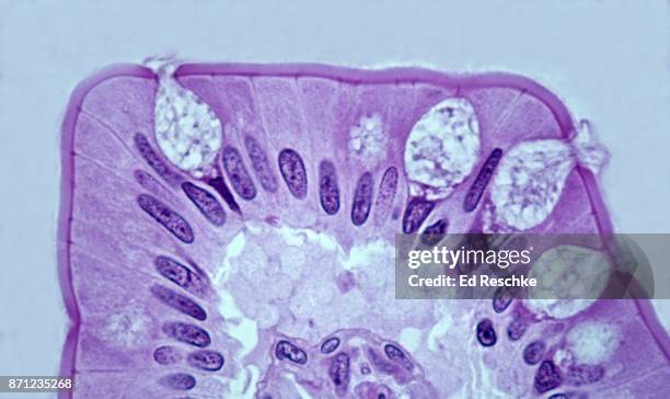 Download premium, authentic SIMPLE COLUMNAR EPITHELIUM and GOBLET CELLS on a Villus in the Small Intestine, 250x stock photos from 51łÔąĎÍř Explore similar high-resolution stock photos in our expansive visual catalogue.Product #:871235268
Download premium, authentic SIMPLE COLUMNAR EPITHELIUM and GOBLET CELLS on a Villus in the Small Intestine, 250x stock photos from 51łÔąĎÍř Explore similar high-resolution stock photos in our expansive visual catalogue.Product #:871235268€300€40
Getty Images
In stockDETAILS
Credit:
51łÔąĎÍř #:
871235268
License type:
Collection:
Stone
Max file size:
5224 x 3104 px (17.41 x 10.35 in) - 300 dpi - 12 MB
Upload date:
Location:
United States
Release info:
No release required
Categories:
- Simple Columnar Epithelial Cell,
- Biology,
- Color Image,
- Desmosome,
- Epithelium,
- Goblet Cell,
- High-scale Magnification,
- Horizontal,
- Human Tissue,
- Lavender Color,
- Macrophotography,
- Magnification,
- Microbiology,
- Microvillus,
- No People,
- Part Of,
- Part of a Series,
- Photography,
- Research,
- Science,
- Scientific Micrograph,
- Small Intestine,
- Technology,
- USA,
- Villus,