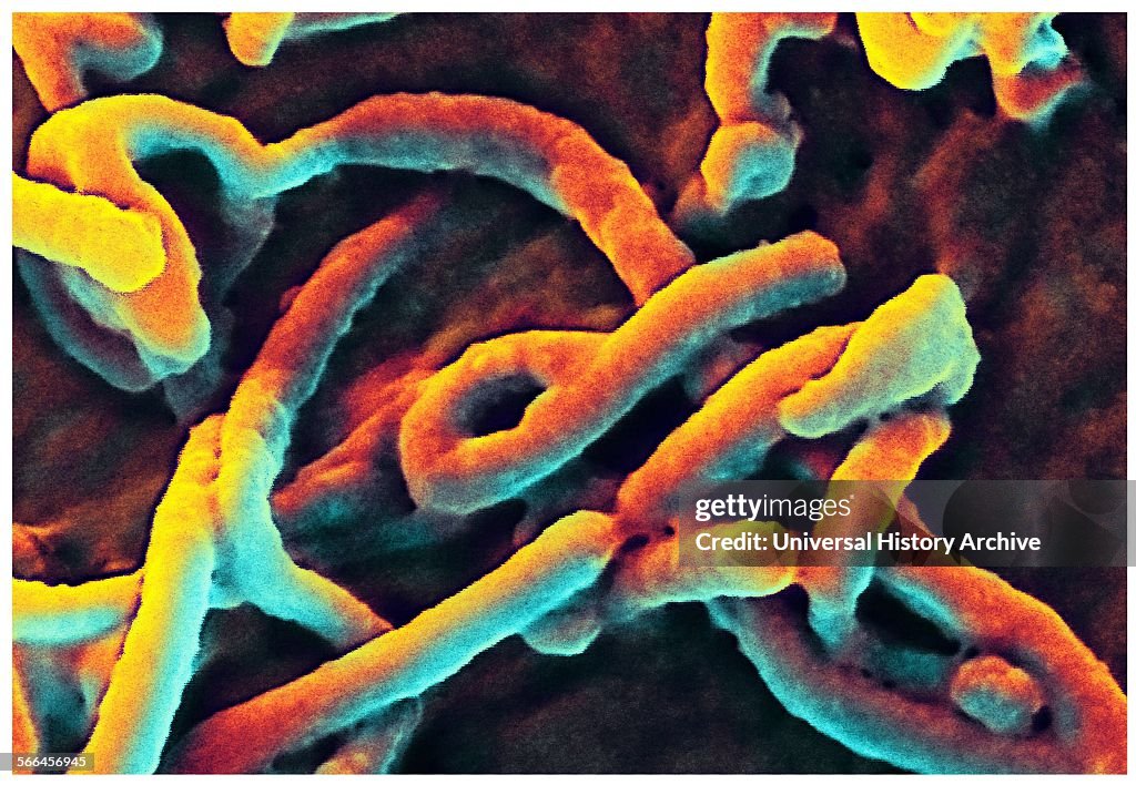
Scanning electron micrograph (SEM) depicts filamentous Ebola virus particles
Produced by the National Institute of Allergy and Infectious Diseases (NIAID), under a very-high magnification, this digitally-colourized scanning electron micrograph (SEM) depicts filamentous Ebola virus particles budding from the surface of a VERO cell of the African green monkey kidney epithelial cell line. (Photo by: Universal History Archive/Universal Images Group via Getty Images)

PURCHASE A LICENSE
How can I use this image?
€300.00
EUR
Getty ImagesScanning electron micrograph (SEM) depicts filamentous Ebola virus..., News Photo Scanning electron micrograph (SEM) depicts filamentous Ebola virus... Get premium, high resolution news photos at Getty ImagesProduct #:566456945
 Scanning electron micrograph (SEM) depicts filamentous Ebola virus... Get premium, high resolution news photos at Getty ImagesProduct #:566456945
€475€115
Getty Images
In stockPlease note: images depicting historical events may contain themes, or have descriptions, that do not reflect current understanding. They are provided in a historical context. .
DETAILS
Restrictions:
Contact your local office for all commercial or promotional uses.
Credit:
Editorial #:
566456945
Collection:
Universal Images Group
Date created:
January 01, 1900
Upload date:
License type:
Release info:
Not released.��More information
Source:
Universal Images Group Editorial
Object name:
917_05_WHA_057_0530
Max file size:
5100 x 3528 px (17.00 x 11.76 in) - 300 dpi - 3 MB