Kidney, Anatomy
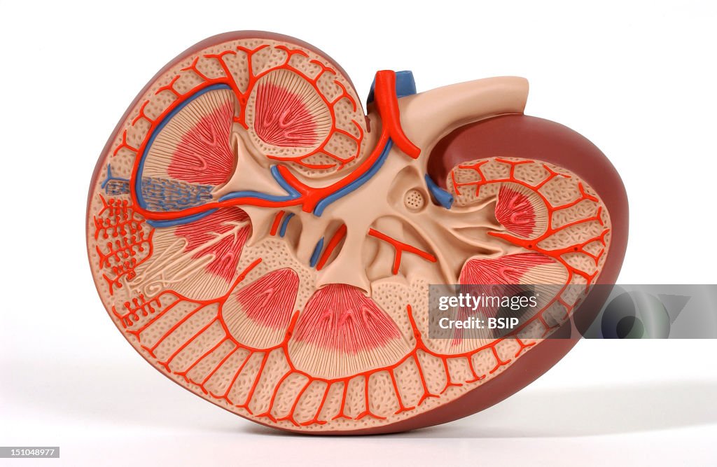
Model Of The Internal Anatomy Of The Right Kidney Of An Adult Human Body Anterior View Of A Frontal Section. The Kidney Filters The Blood, That It Receives At The Hilus Area From The Renal Artery Red. It Makes Urine, Collected In The Renal Pelvis Central Structure Of The Kidney, Hollow, Then Carried By The Ureter Beige Duct To The Urinary Bladder. The Filtered Blood Is Next Drained By The Renal Vein Blue. The Kidney Is Composed Of The Cortex In The Peripheral Area And The Medulla, In The Center, Which Includes Several Malpighian Pyramids Red Separated By The Renal Columns. The Top Of Each Pyramid, Called The Papilla, Leads To A Calyx Hollow That Collects The Urine And Pours It Out Into The Renal Pelvis. In The Cortex And Pyramid Areas, A Multitude Of Nephrons One Is Pictured In Beige On The Left Part Of The Picture Filter The Blood. The Urine Produced By Each Nephron Is Collected By Collecting Tubules And Then Poured Into The Calyces. The Renal Artery Red Branches Into Segmental Then Interlobar Between Each Pyramid And Arcuate Between The Medulla And The Cortex Arteries. The Latter Divide Into Many Interlobular Arterioles Entering The Cortex, That Then Branch Into Afferent Arterioles Shown In The Left Of The Picture. In Parallel, Peritubular Veins Blue Converge In Interlobular, Arcuate, Interlobar, Segmental And Finally Renal Veins. (Photo By BSIP/UIG Via Getty Images)

PURCHASE A LICENSE
How can I use this image?
€300.00
EUR
Getty ImagesKidney, Anatomy, News Photo Kidney, Anatomy Get premium, high resolution news photos at Getty ImagesProduct #:151048977
 Kidney, Anatomy Get premium, high resolution news photos at Getty ImagesProduct #:151048977
€475€115
Getty Images
In stockPlease note: images depicting historical events may contain themes, or have descriptions, that do not reflect current understanding. They are provided in a historical context. .
DETAILS
Restrictions:
Contact your local office for all commercial or promotional uses.
Credit:
Editorial #:
151048977
Collection:
Universal Images Group
Date created:
June 23, 2005
Upload date:
License type:
Release info:
Not released.��More information
Source:
Universal Images Group Editorial
Object name:
941_04_1143805
Max file size:
3630 x 2365 px (12.10 x 7.88 in) - 300 dpi - 2 MB
- Plastic,
- Adult,
- Anatomy,
- Artery,
- Blood,
- Blood Flow,
- Blood Vessel,
- Brain,
- Colored Background,
- Cross Section,
- Front View,
- Horizontal,
- Human Interest,
- Human Internal Organ,
- Human Kidney,
- Kidney Cortex,
- Lumen,
- Medulla,
- Microbiology,
- Nephrology,
- Nephron,
- Papilla,
- People,
- Renal Artery,
- Renal Medulla,
- Renal Pyramid,
- Renal Vein,
- Ureter,
- Urinary System,