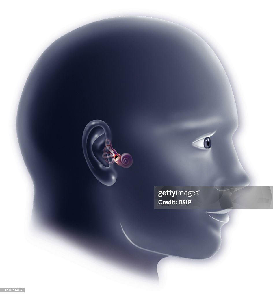
Internal Ear, Drawing
Inner Ear And Vestibular System. The Inner Ear Is The Organ Of Audition And Balance. It Is Composed Of Two Parts The Bony Labyrinth, Externally, That Corresponds To A Series Of Cavities Excavated In The Temporal Bone, And The Membranous Labyrinth, Internally. The Bony Labyrinth Divids Into Three Regions Semi Circular Canals On The Left Of The Image, The Vestibule At The Centre And The Cochlea In Shape Of Snail Shell. The Three Semi Circular Canals Anterior, Posterior And Lateral Are Oriented In The Three Planes Of The Space, Which Enables Them To Perceive The Movements Of The Head And To Participate To Maintain The Dynamic Equilibrium. The Vestibule Includes Two Membranous Sacs, The Utricle And The Saccule Not Represented Here, At The Level Of Which Is Located The Macula In Red Determining The Static Equilibrium. The Cochlea Is A Bony Canal In Shape Of Spiral. It Transforms The Sonor Waves, That Are Perceived By The External Ear And Transmitted By The Middle Ear, In Nerve Impulse. Please Note That The Cochlea Is Overdimensionned Here In Respect To The Reality So That We Can See It Better. (Photo By BSIP/UIG Via Getty Images)

PURCHASE A LICENSE
How can I use this image?
€300.00
EUR
Getty ImagesInternal Ear, Drawing, News Photo Internal Ear, Drawing Get premium, high resolution news photos at Getty ImagesProduct #:151051487
 Internal Ear, Drawing Get premium, high resolution news photos at Getty ImagesProduct #:151051487
€475€115
Getty Images
In stockPlease note: images depicting historical events may contain themes, or have descriptions, that do not reflect current understanding. They are provided in a historical context. .
DETAILS
Restrictions:
Contact your local office for all commercial or promotional uses.
Credit:
Editorial #:
151051487
Collection:
Universal Images Group
Date created:
July 20, 2007
Upload date:
License type:
Release info:
Not released.��More information
Source:
Universal Images Group Editorial
Object name:
941_04_1210407
Max file size:
3351 x 3630 px (11.17 x 12.10 in) - 300 dpi - 936 KB