Pancreatic & Common Bile Ducts
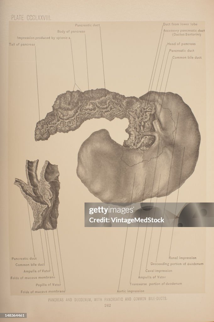
Illustration from 'Surgical Anatomy: The Treatise of the Human Anatomy and Its Applications to the Practice of Medicine and Surgery, volume III' (by Dr. John Blair Deaver) shows the pancreatic duct, a duct joining the pancreas to the common bile duct to supply pancreatic juices which aid in digestion provided by the exocrine pancreas, 1903. The pancreatic duct joins the common bile duct just prior to the ampulla of Vater, after which both ducts perforate the medial side of the second portion of the duodenum at the major duodenal papilla. The common bile ductis a tube-like anatomic structure in the human gastrointestinal tract. It is formed by the union of the common hepatic duct and the cystic duct (from the gall bladder). The duodenum is the first section of the small intestine. It precedes the jejunum and ileum and is the shortest part of the small intestine, where most chemical digestion takes place. The pancreas is a long, slender organ extending form the second portion if the duodenum through the epigastric region, to the hilum of the spleen, in the left hypochondriac region. . (Photo by VintageMedStock/Getty Images)

PURCHASE A LICENSE
How can I use this image?
€300.00
EUR
Getty ImagesPancreatic & Common Bile Ducts, News Photo Pancreatic & Common Bile Ducts Get premium, high resolution news photos at Getty ImagesProduct #:148364461
 Pancreatic & Common Bile Ducts Get premium, high resolution news photos at Getty ImagesProduct #:148364461
€475€115
Getty Images
In stockPlease note: images depicting historical events may contain themes, or have descriptions, that do not reflect current understanding. They are provided in a historical context. .
DETAILS
Restrictions:
Contact your local office for all commercial or promotional uses.
Credit:
Editorial #:
148364461
Collection:
Archive Photos
Date created:
January 01, 1903
Upload date:
License type:
Release info:
Not released.��More information
Source:
Archive Photos
Object name:
T1674624_265
Max file size:
2832 x 4256 px (9.44 x 14.19 in) - 300 dpi - 5 MB