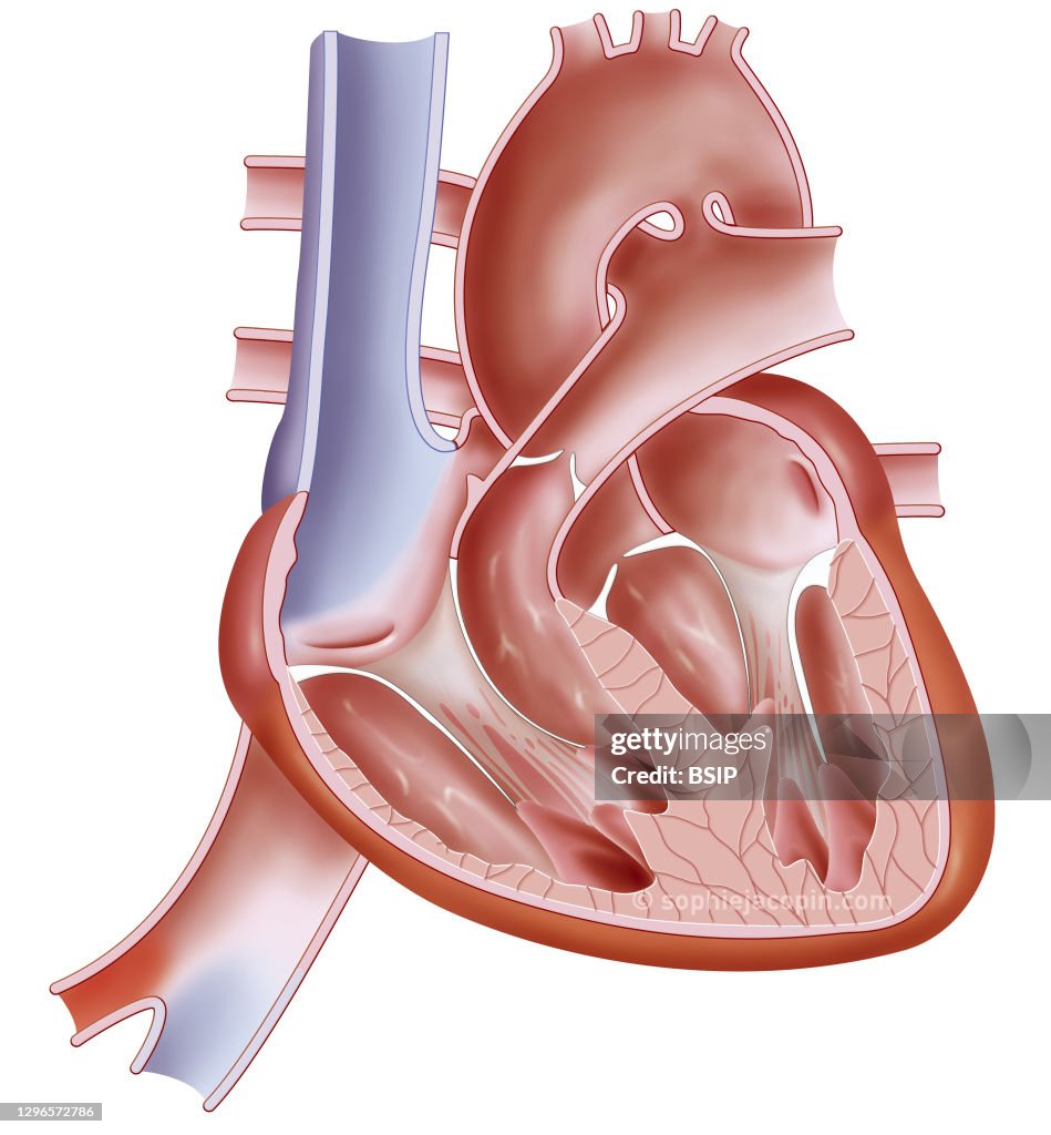
Anatomy of the fetus heart
Heart of a fetus, pediatric anatomy, foramen ovale, arantius venous duct. Illustration representing the heart in a fetus, therefore before birth. Before birth, the right and left atrium communicate through the foramen ovale. This foramen closes at birth due to the pressure caused by the flow of blood. But the aortic arch also communicates with the pulmonary trunk at the level of the arterial duct. This duct closes 3 weeks after birth to become the arterial ligament. Another feature of the fetal heart is an additional blood vessel, the Arantius duct or duct. This vessel is used to transport venous blood from the placenta of the pregnant woman to the inferior vena cava of the fetus without having to pass through the liver. This helps transport oxygen from the umbilical vein much faster. The Arantius canal stops functioning a few minutes after birth. It will spontaneously close again during the first week of life and remain in the form of a residue: the venous ligament of the liver. Blue and red colors indicate oxygenation of the blood. Blue represents deoxygenated blood and red represents oxygenated blood. (Photo by: JACOPIN/BSIP/Universal Images Group via Getty Images)

PURCHASE A LICENSE
How can I use this image?
€300.00
EUR
Getty ImagesAnatomy of the fetus heart, News Photo Anatomy of the fetus heart Get premium, high resolution news photos at Getty ImagesProduct #:1296572786
 Anatomy of the fetus heart Get premium, high resolution news photos at Getty ImagesProduct #:1296572786
€475€115
Getty Images
In stockPlease note: images depicting historical events may contain themes, or have descriptions, that do not reflect current understanding. They are provided in a historical context. .
DETAILS
Restrictions:
Contact your local office for all commercial or promotional uses.
Credit:
Editorial #:
1296572786
Collection:
Universal Images Group
Date created:
November 12, 2020
Upload date:
License type:
Release info:
Not released.��More information
Source:
Universal Images Group Editorial
Object name:
941_04_bsip_016177_024
Max file size:
3795 x 4093 px (12.65 x 13.64 in) - 300 dpi - 4 MB