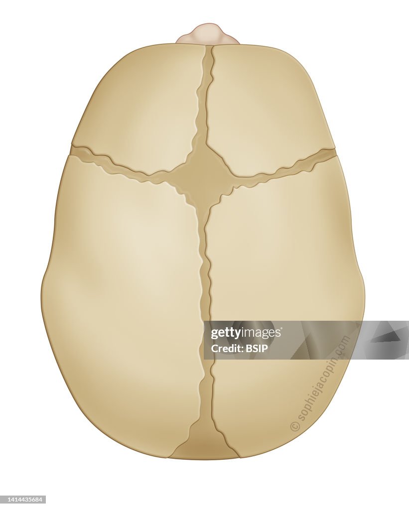
Infant skull - top view
Bone structure at birth, bone development, cartilage structure, pediatrics. Infant skull in top view, fontanel, sutures, pediatrics. This anatomical illustration shows a newborn baby's skull viewed from above. The front of the skull is visible at the top of the drawing thanks to the nose represented. The skull is made up of bony plates, but also of fontanelles and sutures made of connective tissue. These structures give it great elasticity, allowing the baby to pass through the pelvic strait during birth. From top to bottom, we can distinguish the metopic suture, then the anterior fontanel. Coronal sutures are drawn on each side of the anterior fontanel. Then behind the anterior fontanel, it is the sagittal suture. This sagittal suture runs from the anterior fontanel to the posterior fontanel. Two lambdoid sutures, not shown here, start on each side of the posterior fontanel. In the baby, the bones of the skull are not united. This allows the skull to grow according to the rapid growth of the brain during the first years of life. (Photo by: JACOPIN/BSIP/Universal Images Group via Getty Images)

PURCHASE A LICENSE
How can I use this image?
€300.00
EUR
Getty ImagesInfant skull - top view, News Photo Infant skull - top view Get premium, high resolution news photos at Getty ImagesProduct #:1414435684
 Infant skull - top view Get premium, high resolution news photos at Getty ImagesProduct #:1414435684
€475€115
Getty Images
In stockPlease note: images depicting historical events may contain themes, or have descriptions, that do not reflect current understanding. They are provided in a historical context. .
DETAILS
Restrictions:
Contact your local office for all commercial or promotional uses.
Credit:
Editorial #:
1414435684
Collection:
Universal Images Group
Date created:
April 17, 2021
Upload date:
License type:
Release info:
Not released.��More information
Source:
Universal Images Group Editorial
Object name:
941_28_bsip_016251_024
Max file size:
3189 x 3979 px (10.63 x 13.26 in) - 300 dpi - 2 MB