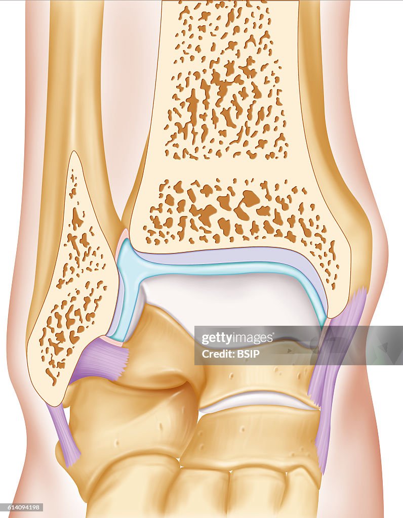
Ankle drawing
Anterior view illustration of the ankle joint. The two joint surfaces (grey) are visible, the joint surface of the fibula and tibia distal epiphysis and that of the talus. The articular capsule is filled with synovial fluid (blue) surrounded by its membrane (pink), it is attached to the lateral ligaments. (Photo by: BSIP/Universal Images Group via Getty Images)

PURCHASE A LICENSE
How can I use this image?
€300.00
EUR
Getty ImagesAnkle drawing, News Photo Ankle drawing Get premium, high resolution news photos at Getty ImagesProduct #:614094198
 Ankle drawing Get premium, high resolution news photos at Getty ImagesProduct #:614094198
€475€115
Getty Images
In stockPlease note: images depicting historical events may contain themes, or have descriptions, that do not reflect current understanding. They are provided in a historical context. .
DETAILS
Restrictions:
Contact your local office for all commercial or promotional uses.
Credit:
Editorial #:
614094198
Collection:
Universal Images Group
Date created:
April 01, 2016
Upload date:
License type:
Release info:
Not released.��More information
Source:
Universal Images Group Editorial
Object name:
941_28_bsip_014968_013.jpg
Max file size:
3950 x 5079 px (13.17 x 16.93 in) - 300 dpi - 5 MB
- Anatomy,
- Ankle,
- Art Product,
- Articular Capsule,
- Astragalus,
- Bone,
- Cartilage,
- Collateral Ligament,
- Discussion,
- Foot,
- France,
- Front View,
- Human Interest,
- Human Skeleton,
- Lateral Collateral Ligament,
- Leg,
- Ligament,
- Limb - Body Part,
- Medial Collateral Ligament,
- Medicine,
- Navicular Bone Of Hand,
- No People,
- Orthopedics,
- People,
- Photography,
- Rheumatology,
- Talus,
- Tarsus,
- Tibia,
- Vertical,