Testicle anatomy, illustration - stock illustration
Illustration of the anatomy of the human testis (testicle), part of the male reproductive system. The testes are where the male sex cells (spermatozoa) are produced. This process, called spermatogenesis, occurs in the seminiferous tubules, within the main body of the testicle. To the left of the testicle is the epididymis, which carries the sperm to the spermatic duct (vas deferens)

Get this image in a variety of framing options at Photos.com.
PURCHASE A LICENSE
All Royalty-Free licenses include global use rights, comprehensive protection, simple pricing with volume discounts available
€300.00
EUR
Getty ImagesTesticle Anatomy Illustration High-Res Vector Graphic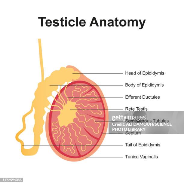 Download premium, authentic Testicle anatomy, illustration stock illustrations from 51łÔąĎÍř Explore similar high-resolution stock illustrations in our expansive visual catalogue.Product #:1472594088
Download premium, authentic Testicle anatomy, illustration stock illustrations from 51łÔąĎÍř Explore similar high-resolution stock illustrations in our expansive visual catalogue.Product #:1472594088€300€40
Getty Images
In stockDETAILS
51łÔąĎÍř #:
1472594088
License type:
Collection:
Science Photo Library
Max file size:
4968 x 4968 px (16.56 x 16.56 in) - 300 dpi - 2 MB
Upload date:
Release info:
No release required
Categories: