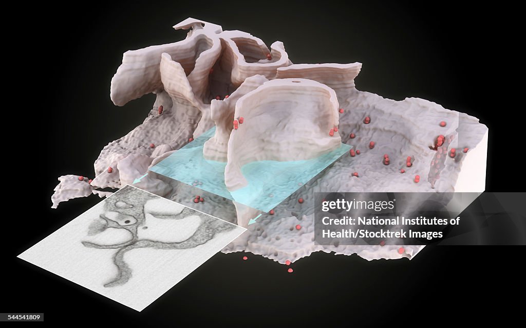
Surface of HIV infected microphage. - stock illustration
3D representation of the surface and interior of an HIV-infected macrophage obtained using newly developed tools for 3D imaging using ion-abrasion scanning electron microscopy. Sections that would appear to contain filopodia when imaged by transmission electron microscopy of individual sections can actually correspond to large wavelike membrane processes as illustrated by the cut-away view of the slice. These surface protrusions may potentially fold back to the surface of the cell, creating viral compartments (viruses shown in red) by trapping the contents of the aqueous environment within the invaginated folds of the membrane.

Get this image in a variety of framing options at Photos.com.
PURCHASE A LICENSE
All Royalty-Free licenses include global use rights, comprehensive protection, simple pricing with volume discounts available
€300.00
EUR
Getty ImagesSurface Of Hiv Infected Microphage High-Res Vector Graphic Download premium, authentic Surface of HIV infected microphage. stock illustrations from 51łÔąĎÍř Explore similar high-resolution stock illustrations in our expansive visual catalogue.Product #:544541809
 Download premium, authentic Surface of HIV infected microphage. stock illustrations from 51łÔąĎÍř Explore similar high-resolution stock illustrations in our expansive visual catalogue.Product #:544541809
€300€40
Getty Images
In stockDETAILS
51łÔąĎÍř #:
544541809
License type:
Collection:
Stocktrek Images
Max file size:
4050 x 2526 px (13.50 x 8.42 in) - 300 dpi - 1 MB
Upload date:
Release info:
No release required
Categories:
- Filopodium,
- Analyzing,
- Bacterium,
- Biological Cell,
- Biology,
- Biotechnology,
- Black Background,
- Blood,
- Cell Membrane,
- Close-up,
- Color Image,
- Cross Section,
- Cutaway Drawing,
- Cytoplasm,
- Electron Micrograph,
- Growth,
- HIV,
- Healthcare And Medicine,
- Horizontal,
- Human Tissue,
- Illustration,
- Indoors,
- Infectious Disease,
- Macrophage,
- Magnification,
- Membrane,
- Microbiology,
- No People,
- Pathogen,
- Research,
- Science,
- Scientific Micrograph,
- Three Dimensional,
- Trapped,
- Virus,