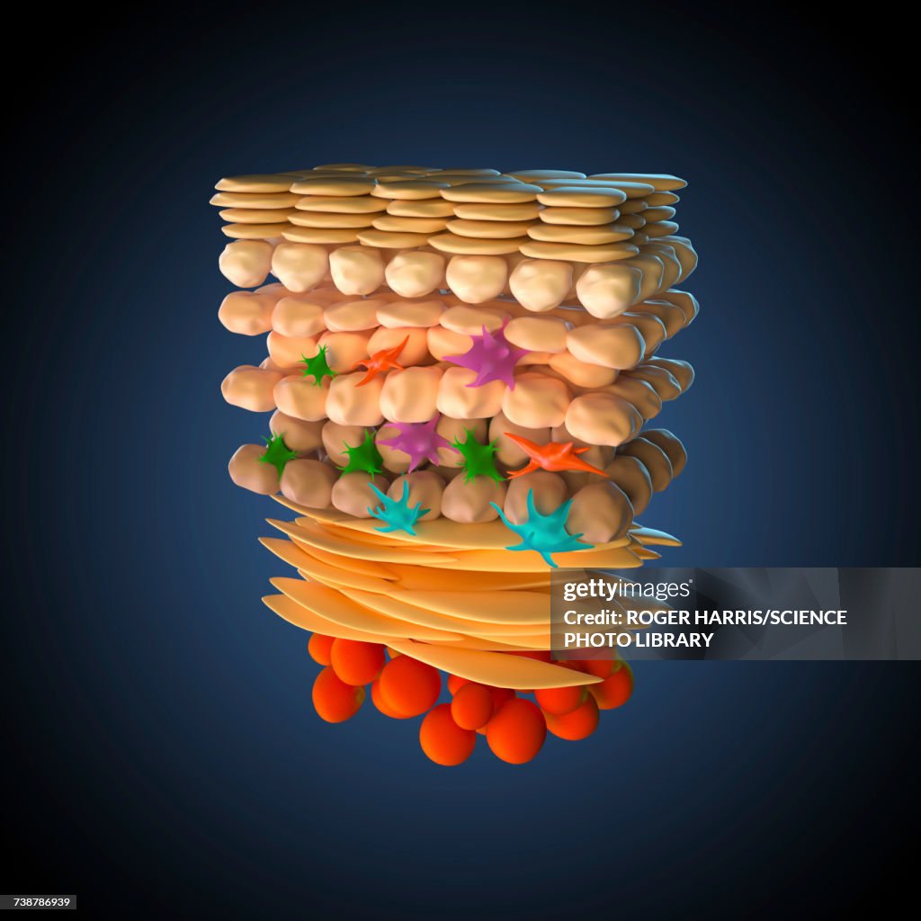
Skin anatomy, illustration - stock illustration
Illustration of a cross-section through human skin. At bottom are adipocyte (fat) cells (red). Above this is the dermis (flattened yellow cells), and then the basal, spinous and granular layers of the epidermis and keratinised dead cells at the surface. Also shown are fibroblasts (orange), melanocytes (green), langerhans cells (pink) and merkel cells (blue).

Get this image in a variety of framing options at Photos.com.
PURCHASE A LICENSE
All Royalty-Free licenses include global use rights, comprehensive protection, simple pricing with volume discounts available
€300.00
EUR
Getty ImagesSkin Anatomy Illustration High-Res Vector Graphic Download premium, authentic Skin anatomy, illustration stock illustrations from 51łÔąĎÍř Explore similar high-resolution stock illustrations in our expansive visual catalogue.Product #:738786939
 Download premium, authentic Skin anatomy, illustration stock illustrations from 51łÔąĎÍř Explore similar high-resolution stock illustrations in our expansive visual catalogue.Product #:738786939
€300€40
Getty Images
In stockDETAILS
51łÔąĎÍř #:
738786939
License type:
Collection:
Science Photo Library
Max file size:
5100 x 5100 px (17.00 x 17.00 in) - 300 dpi - 5 MB
Upload date:
Release info:
No release required
Categories:
- Skin,
- Science,
- Anatomy,
- Cross Section,
- Illustration,
- Adipose Cell,
- Basal Cell,
- Human Skin,
- Islet of Langerhans,
- Three Dimensional,
- Art Product,
- Biology,
- Biomedical Illustration,
- Built Structure,
- Color Image,
- Dermis,
- Digitally Generated Image,
- Fibroblast,
- Healthcare And Medicine,
- Human Body Part,
- Human Digestive System,
- Human Internal Organ,
- Melanocyte,
- No People,
- Square - Composition,