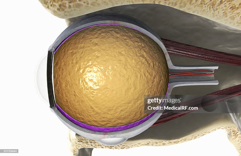
Lateral view of the right eyeball exposed in the eye socket. - stock illustration
This image is a lateral view of the right eyeball exposed in a cutaway section of the eye socket, showing the superior rectus muscle and the inferior rectus muscle. The optical nerve is also shown and its initial route through the skull. The sclera and choroid layers have also been removed from one half of the eyeball.

Get this image in a variety of framing options at Photos.com.
PURCHASE A LICENSE
All Royalty-Free licenses include global use rights, comprehensive protection, simple pricing with volume discounts available
€300.00
EUR
Getty ImagesLateral View Of The Right Eyeball Exposed In The Eye Socket High-Res Vector Graphic Download premium, authentic Lateral view of the right eyeball exposed in the eye socket. stock illustrations from 51łÔąĎÍř Explore similar high-resolution stock illustrations in our expansive visual catalogue.Product #:55733664
 Download premium, authentic Lateral view of the right eyeball exposed in the eye socket. stock illustrations from 51łÔąĎÍř Explore similar high-resolution stock illustrations in our expansive visual catalogue.Product #:55733664
€300€40
Getty Images
In stockDETAILS
Credit:
51łÔąĎÍř #:
55733664
License type:
Collection:
MedicalRF.com
Max file size:
5100 x 3300 px (17.00 x 11.00 in) - 300 dpi - 3 MB
Upload date:
Release info:
No release required
Categories:
- Anatomy,
- Biomedical Illustration,
- Bulbar Conjunctiva,
- Choroid,
- Close-up,
- Color Image,
- Digitally Generated Image,
- Eye Socket,
- Eyeball,
- Horizontal,
- Human Body Part,
- Human Internal Organ,
- Human Skull,
- Illustration,
- Illustration Technique,
- Inferior Oblique Muscle,
- Inferior Rectus Muscle,
- Lateral Rectus Muscle,
- No People,
- Optometry,
- Physiology,
- Retina,
- Sclera,
- Side View,
- Studio Shot,
- Superior Rectus Muscle,
- White Background,