Diatoms and radiolaria, SEM - stock illustration
Diatoms and radiolaria. Coloured scanning electron micrograph (SEM) of a circular arrangement of various diatoms and radiolaria. Diatoms are planktonic unicellular algae. They have a mineralised cell wall (frustule) divided into two halves. The frustule contains silica and provides protection and support. Radiolarians are amoeboid protozoa that produce intricate mineral skeletons. They are found as zooplankton throughout the ocean, and their skeletal remains cover large portions of the ocean bottom as radiolarian ooze.

Get this image in a variety of framing options at Photos.com.
PURCHASE A LICENSE
All Royalty-Free licenses include global use rights, comprehensive protection, simple pricing with volume discounts available
€300.00
EUR
Getty ImagesDiatoms And Radiolaria Sem High-Res Vector Graphic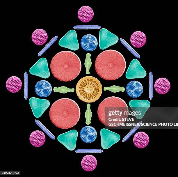 Download premium, authentic Diatoms and radiolaria, SEM stock illustrations from 51łÔąĎÍř Explore similar high-resolution stock illustrations in our expansive visual catalogue.Product #:685023393
Download premium, authentic Diatoms and radiolaria, SEM stock illustrations from 51łÔąĎÍř Explore similar high-resolution stock illustrations in our expansive visual catalogue.Product #:685023393€300€40
Getty Images
In stockDETAILS
51łÔąĎÍř #:
685023393
License type:
Collection:
Science Photo Library
Max file size:
4572 x 4539 px (15.24 x 15.13 in) - 300 dpi - 6 MB
Upload date:
Release info:
No release required
Categories:
- Diatom,
- Abdomen,
- Algae,
- Anatomy,
- Animal Digestive System,
- Animal Esophagus,
- Art Product,
- Biology,
- Biomedical Illustration,
- Black Background,
- Blue Background,
- Color Image,
- Colored Background,
- Digestive System,
- Digitally Generated Image,
- Esophagus,
- Front View,
- Healthcare And Medicine,
- Horizontal,
- Human Internal Organ,
- Illustration,
- Internal Organ,
- No People,
- Protozoan,
- Radiolaria,
- SEM,
- Science,
- Three Dimensional,
- Unicellular Organism,